Nankai Research Team Achieved Photooxidation-Driven Purely Organic Room-Temperature Phosphorescent Lysosome-Targeted Imaging
Recently, Professor Liu Yu’s Research Group from College of Chemistry, Nankai University, found that the photooxidation-driven purely organic room-temperature phosphorescent supramolecular system can achieve dual targeted locating and imaging of cell nucleus and lysosome, which greatly expands the application scope of the supramolecular system.
Related research results were published on Journal of the American Chemical Society on August 19, 2021 with the title of “Photooxidation-Driven Purely Organic Room-Temperature Phosphorescent Lysosome-Targeted Imaging”. Professor Liu Yu and Associate Professor Zhang Yingming are the joint corresponding authors, and Yu Huajiang, a doctoral student from College of Chemistry, is the first author.
Based on the previous work, the research group skillfully designed and synthesized a photooxidation-driven supramolecular fluorescent and phosphorescent bidirectional luminescence system and successfully achieved multi-color targeted imaging for double organelles in living cells. Based on the molecular recognition of cucurbituril (a kind of macrocyclic compound condensed by glycoluril and formaldehyde) and photooxidation of bound guest anthracene, they found that the macrocyclic compound can effectively induce the assembly of anthracyl conjugate bromophenyl pyridine salts to form supramolecular polymer through the stable intermolecular charge transfer interaction of cucurbituril, which led to an increase in rigidity and conjugate degree, thus significantly improving the intensity of red fluorescence emission. In contrast, under light conditions, the anthryl is oxidized to anthraquinone and the molecular assembly pattern is also transformed from the “head to tail” supramolecular polymer to the “head to head” ternary compound, and the ternary compound can emit strong green phosphorescence in aqueous solution.
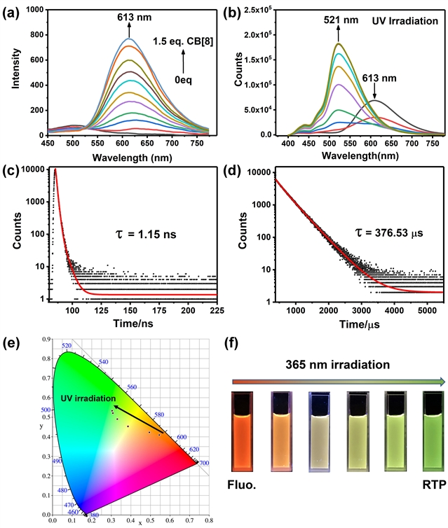
Figure 1: Spectral Characterization of Assembly Aqueous Solution.(a) Fluorescent spectral changes in supramolecular polymer formed by adding cucurbituril [8] into guest. (b) Photoluminescent spectral changes in supramolecular polymer under UV irradiation. (c) Life of supramolecular polymer at 613nm under room temperature. (d) Life of ternary compound formed after illumination at 529nm under room temperature. (e) CIE 1931 chromaticity diagram of supramolecular polymer with UV irradiation.(f) Luminescence photo of supramolecules under continuous UV light.
Furthermore, the authors have successfully transformed and applied the photophysical properties of fluorescent-phosphorescent emission to targeted imaging of different organelles in living cells. The experimental results of cell staining show that the supramolecular polymer with red fluorescence emission ability can be well located and imaged in cell nucleus, while supramolecular ternary compound with green phosphorescence emission ability can be selectively located in lysosome. In addition, after cell co-incubation of supramolecular polymer and lysosomal imaging agent, it was found that supramolecular polymer can be recombined into ternary compound in cells under light conditions, and is transformed into green fluorescence in lysosome with the red fluorescence in cell nucleus.
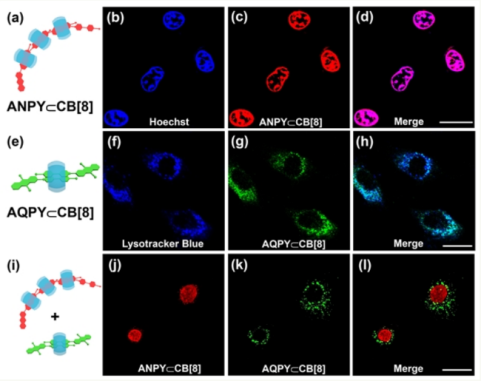
Figure 2: Experimental Results of Cell Staining in Supramolecular Assembly.(a-d) Supramolecular polymers, (e-h) Supramolecular ternary compounds, (i-l) Cell imaging maps of two assemblies.
Researchers held that the work has achieved multi-color targeted imaging for different organelles in living cells for the first time by means of photooxidation-driven strategy, which provides a new idea for developing stimuli-responsive materials and functional imaging reagents.
Link to the paper: https://doi.org/10.1021/jacs.1c06741
38 Tongyan Road, Jinnan District, Tianjin, P.R.China 300350 94 Weijin Road, Nankai District, Tianjin, P.R.China 300071
Copyright: Nankai University All Rights Reserved