NKU Team Developed U-ExSMLM to Reveal the Asymmetry of Cytoskeleton in Biconcave Erythrocytes

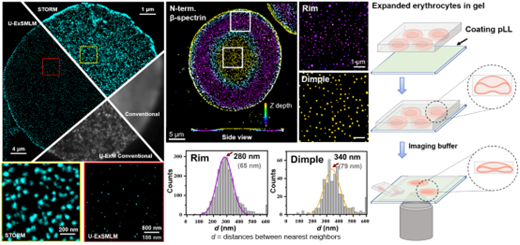
Recently, a Nankai University team led by Professor Pan Leiting and Professor Xu Jingjun of the School of Physics and TEDA Institute of Applied Physics developed super-resolution imaging technology (U-ExSMLM) based on ultrastructure expansion microscopy (U-ExM) combined with single-molecule localization microscopy (SMLM), reaching a molecular resolution of about 6 nm. At the same time, the team studied human erythrocyte cytoskeletal ultrastructure under near-physiological conditions, and intuitively revealed the asymmetrical distribution of cytoskeleton in biconcave erythrocytes, providing a powerful molecular-level imaging explanation for learning about the unique biconcave shape and shape memory of erythrocyte. The results entitled “Molecular Resolution Mapping of Erythrocyte Cytoskeleton by Ultrastructure Expansion Single-Molecule Localization Microscopy (U-ExSMLM)” were published in Small Methods, a publication run by Wiley Group.
Skillfully adhering the gel onto poly-lysine-coated coverslip to prevent lateral shrinkage of the hydrogel, the team successfully achieved the combination of 4.3-fold physically expanded ExM and SMLM, improving the 25 nm resolution of SMLM to a molecular resolution level of about 6 nm.
The team developed the weak adhesion conditions that can maintain the biconcave shape of erythrocytes, and performed imaging with U-ExSMLM. It was found that the spectrin cytoskeleton in the dimple regions has lower density and larger length than that in the rim regions. It provides a novel super-resolution imaging method for studying the subcellular structure and functions near cytoplasmic membrane.
The team led by Professor Pan Leiting and Professor Xu Jingjun has long been committed to studying the development and application of SMLM, and has made a series of achievements. For instance, based on SMLM, the team has figured out the physiological length of erythrocyte spectrin, a question that has existed for 40 years (Cell Reports, 2018), revealed the rapid and reversible depolymerization and assembly characteristics of intermediate filaments in response to hypotonic stimulation at the nanoscale (Advanced Science, 2019), and found that the three-dimensional architecture of macrophage podosome resembles a “snowman”-like rather than the traditionally believed “dome”-like portrait (iScience, 2022).
The co-first authors of this paper are doctoral student Hou Mengdi and postdoctoral fellow Xing Fulin, the corresponding author is Professor Pan Leiting, while Nankai University is the first author’s affiliation.
Link to the paper: https://onlinelibrary.wiley.com/doi/full/10.1002/smtd.202201243


