NKU Team Established High-Throughput and High-Content Drug Screening Method Based on Microarray and Expansion Microscopy
Combination of high-throughput microarray technology and high-content expansion microscopy (ExM)
The latest research in the field of drug screening conducted by Prof. Qing Ye and Prof. Jianguo Tian from the School of Physics of Nankai University was published in ACS Nano, a world-renowned academic journal in the field of materials. They established an imaging-based high-throughput and high-content drug screening method by combining microarray preparation technology with expansion microscopy (ExM). Compared with existing technologies, this method improves throughput and resolution simutaneously, while slashing time and material costs. More over, this method is easy to implement and available in any biomedical laboratory, regardless of the expensive equipment and the experienced technician. The accessibility and high efficiency of this method will provide a powerful methodology for image-based drug screening, especially for the testing of some ultrastructure-targeted drugs, which contributes greatly to the early screening of drugs.
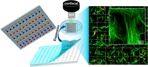
The research group provided a drug screening method exhibiting high throughput and high content simutaneously. First of all, superhydrophobic microwell array plate (SMAP) was prepared based on photolithography technology. Compared with traditional multiwell plates, the miniaturized and highly integrated SMAP greatly improves the detection throughput. when the cells reaches about 80% confluency, the microcolumn–microwell sandwiching technology was applied to realize drug releasing. This method can achieve parallel drug treatment in a time-saving manner, dispensing with the use of expensive microarrayer. After drug treatment, ExM was applied and the fluorescence images of microtubules and mitochondria of COS-7 and HeLa cells were collected using confocal microscopy. Afterwards, they carried out qualitative and quantitative analysis of the morphological changes of microtubules and mitochondria, which demonstrated that the application of ExM greatly improves the imaging resolution and provides more details on the morphological changes of cell submicroscopic structure than traditional diffraction-limit microscopy. This high-throughput and high-content drug screening method offers an alternative technical means for the study of pharmacologic action.
Junfang Xie, a doctoral student, is the first author of the paper.
Link:https://pubs.acs.org/doi/10.1021/acsnano.3c01865
(Edited and translated by Nankai News Team.)
38 Tongyan Road, Jinnan District, Tianjin, P.R.China 300350 94 Weijin Road, Nankai District, Tianjin, P.R.China 300071
Copyright: Nankai University All Rights Reserved